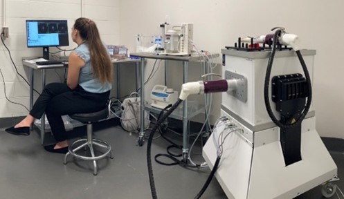
Aspect Imaging M3™ Compact MRI System Information

The System generates high resolution 2D and 3D anatomical, functional, and molecular images using a bore size optimized for laboratory mice. This 1-T preclinical MRI system allows for studying disease progression and biodistribution of biological active molecules using targeted contrast agents such as gadolinium and superparamagnetic iron oxide nanoparticles and other metal ions chelated with antibodies. The system consists of a fully integrated animal handling system that has two designated RF coils for different imaging applications. One RF coil (FOV: 23 x 25mm) is used for neurological imaging of the head, while the second (FOV: 30 x 50mm) is used for extremity, abdominal and thoracic cavity imaging in mice. The system monitors respiration, ECG, and temperature while the laboratory animals are being imaged under isoflurane-based anesthesia. VivoQuant™ and M-Series™ imaging software allow post processing of MR imaging data such as fine-tuning images, isolating and analyzing regions of interest and allow 3D MIP and volume.
Please see the Animal Research Facility section for information on receiving access to the facility and equipment.
