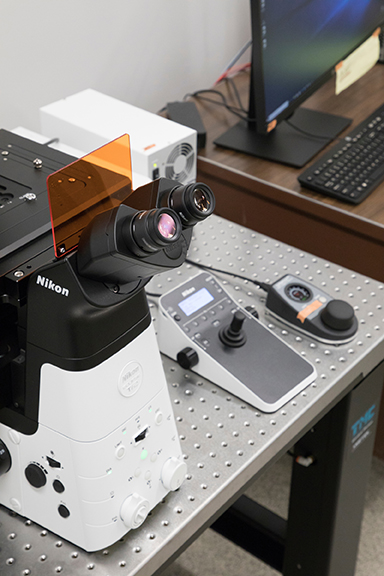
Nikon A1R-HD Confocal Microscope (Ti2) Information

The Nikon A1R Confocal Microscope is a powerful, fully-automated confocal imaging system. The system is able to image in the X, Y, and Z planes with both a high-speed resonant scanner to capture the dynamics of a system and a Galvano scanner to maximize resolution in fixed samples. The A1R-HD a large field of view and is capable of a resolution below 200 nm. The system comes with sophisticated post-processing software to deconvolve and attractively display results. The system has a 2D and 3D tracking system for particle tracking can be set up to automatically track several regions of interest in a time series scan.
The system includes the following subsystems and components:
- Nikon A1R Live Cell Resonant Dual Scanner
- A1-DUG Hybrid GaAsP/PMT 4ch Detector System
- Transmitted Light Detector
- Nikon LUN4 4 Line Solid State Laser System
- Ti2e Fully Motorized Inverted Microscope
- Nikon Perfect Focus System
- Motorized Encoded XY Stage
- LED Based Epi Fluorescent System (DAPI, FITC, TRITC cubes)
- LED Based Transmitted Light System
- Lasers in the blue, green, red and far-red channels
- 10x, 20x, 40x oil, 60x oil Objectives
- 20x long-working distance objective for live-cell and dynamics imaging
- DIC Imaging
- Stage-Top Incubator for Temperature and Carbon Dioxide Control
- Deconvolution software for improved visualization
- Dedicated analysis workstation for processing images and making presentation quality images
- Spectral Imager capable of 2.5 nm wavelength resolution and capture of 32 channels fluorescent spectra in a a single scan
- Elimination of autofluorescence and separation of Fluorophores with overlapping spectra
For training, please contact Katie Shannon at shannonk@mst.edu or 573-341-6336.
Objective Information
| Magnification | Numerical Apeture | Working Distance | Immersion Medium | Optical Corrections |
|---|---|---|---|---|
| 10x | .45 NA | 4.0 mm | air | Plan Apo |
| 20x | .75 NA | 1.0 mm | air | Plan Apo VC |
| 20x | .95 NA | .95 mm | water | CFI Apo LWD Lambda S |
| 40x | 1.30 NA | 0.20 mm | oil | Plan Fluor |
| 60x | 1.40 NA | 0.13 mm | oil | Plan Apo Lambda |
All objectives configured for #1.5 coverslips, 20x water and 40x oil objectives also have correction collars for plastic culture dishes. Please see MicroscopyU for more information.
Confocal Laser Lines
| Laser line for excitation | Emission Filter | Example Fluorophore |
|---|---|---|
| 405 nm | 425-475 nm | DAPI, CFP |
| 488 nm | 500-550 nm | FITC, GFP, Alexa 488 |
| 561 nm | 570-620 nm | TRITC, Alexa 568, Texas Red, Pl |
| 640 nm | 633-673 nm | Cy5 |
A useful website to check compatibility of your fluorophore with the laser lines: https://www.thermofisher.com/us/en/home/life-science/cell-analysis/labeling-chemistry/fluorescence-spectraviewer.html
